The skin, being the largest organ, acts as a primary interface between the body’s internal environment and the external world. Consequently, it is often the first to exhibit distress in the form of various dermatologic eruptions. A person noticing an area of red, inflamed, or itchy skin frequently defaults to the term “rash,” a broad and non-specific descriptor encompassing hundreds of possible causes. However, correctly identifying the underlying mechanism—whether it is a simple, localized irritation (a rash) or a more complex, systemic immunological response (an allergic reaction)—is critical for effective treatment and management. Misdiagnosis can lead to the inappropriate use of medications, delayed resolution, or, in severe cases, life-threatening complications. The distinction is not always straightforward, as both categories share overlapping visual characteristics like redness, swelling, and pruritus (itching). Navigating this diagnostic challenge requires a deeper understanding of the immunological pathways and external triggers involved, moving beyond superficial appearances to understand the kinetic and morphological clues that point to the true origin of the skin’s distress.
The Skin Is Often the First to Exhibit Distress in the Form of Various Dermatologic Eruptions
A key differentiator lies in the mechanism of injury and the immune pathways activated. A simple irritant contact dermatitis, the most common type of non-allergic rash, occurs when the skin is physically or chemically damaged by a substance. This direct toxicity requires no prior sensitization. For instance, prolonged exposure to harsh cleaning agents, solvents, or even excessive friction can strip the skin’s protective barrier, leading to immediate inflammation. The resulting rash is typically confined precisely to the area of contact and often presents as dry, cracked, and sometimes painful skin. This reaction is a non-immunological, dose-dependent response: the harsher the chemical or the longer the exposure, the more severe the reaction will be. In contrast, an allergic contact dermatitis is an immune-mediated, hypersensitivity response. It requires a specific antigen (allergen) to trigger a delayed type IV hypersensitivity reaction, meaning the immune system has been sensitized in the past, and the reaction only appears hours or days after re-exposure.
An Allergic Contact Dermatitis Is an Immune-Mediated, Hypersensitivity Response
The characteristic presentation of an allergic contact dermatitis is an immune-mediated, hypersensitivity response, distinguishing it visually from simple irritation. Because it is driven by the migration of T-cells to the site of contact, the resulting inflammation often extends slightly beyond the initial exposure area and can be highly vesicular (blistering) or intensely itchy. Classic examples involve exposure to poison ivy/oak (urushiol) or common cosmetic ingredients like certain fragrances or preservatives. The pattern of the rash can be highly instructive; a reaction to a watch strap might form a clear band on the wrist, while an allergy to a nail polish might manifest unexpectedly on the eyelids or neck, areas the individual inadvertently touched after applying the polish. Furthermore, the allergic reaction is not dose-dependent in the same way; even trace amounts of the allergen can elicit a severe, widespread response in a sensitized individual, highlighting the profound difference in the underlying biological pathway compared to simple irritant rashes.
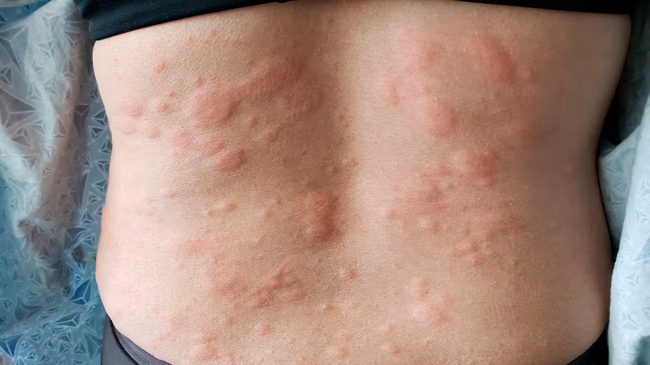
A Drug Eruption is a Systemic Response, Often Bilateral and Symmetrical
When the body reacts to an ingested or injected substance, such as a medication, the resulting skin issue is termed a drug eruption. This differs from contact dermatitis because the allergen enters the bloodstream and circulates throughout the body. Therefore, a drug eruption is a systemic response, often bilateral and symmetrical, appearing simultaneously across large body areas like the trunk, arms, and legs. Drug reactions can manifest in dozens of ways, from a simple morbilliform (measles-like) rash to potentially fatal conditions like Stevens-Johnson Syndrome/Toxic Epidermal Necrolysis (SJS/TEN). The timing is a vital diagnostic clue; while some drug allergies appear quickly (within hours), others, particularly those mediated by T-cells, can take days or even weeks to emerge after starting a new medication. Since the skin is merely the target organ for a systemic immunological cascade, stopping the offending medication is the immediate, non-negotiable step in management, followed by supportive care to manage the widespread inflammation.
The Immediate Appearance of Urticaria is Mediated by Mast Cells
Another dramatic and immediately recognizable form of allergic reaction is urticaria, commonly known as hives. The immediate appearance of urticaria is mediated by mast cells and represents a type I hypersensitivity reaction. Unlike the delayed T-cell response, this reaction is rapid, often occurring within minutes of exposure to an allergen (e.g., peanuts, penicillin, bee venom). Hives present as intensely itchy, raised wheals (welts) that are transient—they can appear, swell, and fade within hours, often changing location across the body. This ephemeral nature is a hallmark of urticaria and is due to the release of histamine and other potent mediators from mast cells, causing localized vasodilation and fluid leakage into the skin. While often a benign annoyance, urticaria can be the initial sign of anaphylaxis, especially if accompanied by difficulty breathing, throat swelling, or a drop in blood pressure, necessitating immediate emergency medical intervention.
Environmental Factors Can Also Play a Crucial, Confusing Role
The diagnostic process is further complicated by the interaction of external factors and the patient’s internal state. Environmental factors can also play a crucial, confusing role by acting as co-factors that precipitate or exacerbate both allergic and non-allergic skin conditions. For instance, exposure to sunlight combined with certain medications (a photoallergic or phototoxic reaction) can produce a severe, rash-like burn on sun-exposed areas. Similarly, the presence of heat and sweating can aggravate underlying conditions like atopic dermatitis (eczema), making it appear much worse and more inflammatory. A dermatologist must meticulously deconstruct the timeline, location, and morphology of the eruption, cross-referencing these clues with the patient’s complete exposure history—including all cleaning products, clothing, hobbies, and medications—to filter out these confounding variables and isolate the true primary trigger, whether it’s an allergen or an irritant.
The Primary Tool is a Detailed and Comprehensive Patient History
Ultimately, discerning the difference between an irritant rash and a true allergic reaction in clinical practice rarely relies on a single test. Instead, the primary tool is a detailed and comprehensive patient history, sometimes extending back over the patient’s entire lifetime. This historical interrogation seeks to identify the pattern of recurrence, the exact timeline from exposure to symptom onset, the precise location of the initial lesion, and whether the rash is migratory or fixed. For contact allergies, the key is the spatial relationship between the rash and a potential allergen. For systemic drug reactions, the key is the temporal relationship between starting the medication and the rash’s appearance. Laboratory tests, such as IgE blood tests for immediate allergies or patch testing for delayed contact allergies, serve only to confirm a suspicion generated by the careful analysis of the patient’s narrative, proving that clinical detective work often precedes molecular confirmation.
Patch Testing Serves as the Gold Standard for Contact Allergies
For diagnosing delayed-type contact hypersensitivity, patch testing serves as the gold standard for contact allergies. This procedure involves applying small quantities of common potential allergens (a standard battery of 30-80 substances, ranging from metals like nickel to chemicals found in rubber, leather, or cosmetics) to the patient’s back under occlusive patches. These patches remain in place for 48 hours, and the test sites are evaluated at 48 hours and again at 72 or 96 hours. A positive result—manifesting as localized redness, papules, or vesicles precisely where the allergen was placed—provides irrefutable evidence of a type IV cell-mediated immunity to that specific substance. Unlike prick or IgE tests, which look for immediate, histamine-driven allergies, patch testing directly recreates the delayed immune response, offering the crucial diagnostic information necessary for the patient to permanently avoid the trigger and achieve clearance of their chronic dermatitis.
Complete Avoidance of the Identified Trigger Is the Curative Strategy
Once a true contact allergy has been identified through meticulous history and confirmed via patch testing, complete avoidance of the identified trigger is the curative strategy. Because the immune system’s memory of the allergen is often lifelong, re-exposure, even years later, will provoke the reaction again. This is where patient education becomes paramount, requiring patients to read ingredient lists on all products they use—from shampoos and sunscreens to workplace chemicals and clothing dyes. Avoidance is the most effective and least pharmacologic treatment available. For non-allergic irritant dermatitis, the strategy is less about lifelong avoidance and more about barrier protection (using gloves) and repairing the skin’s natural barrier function with emollients and thick creams. The difference in treatment approaches perfectly reflects the difference in pathology: resolving inflammation versus rewriting an immune memory.
Inflammation of the Vasculature Can Cause Palpable Purpura
Beyond typical rashes and urticaria, certain systemic conditions can present with skin manifestations that are the result of damage to the blood vessels themselves. Inflammation of the vasculature can cause palpable purpura, a term used to describe red or purple spots on the skin that do not fade when pressed (non-blanching). This is often a sign of vasculitis, where immune complexes or direct immune attack cause inflammation and leakage of red blood cells from the small vessels, a condition that can be triggered by drugs, infections, or underlying autoimmune diseases. This type of skin lesion is a profound red flag, indicating a systemic process that can affect internal organs like the kidneys, lungs, or gut. Palpable purpura is a dermatologic emergency that always warrants immediate medical workup, as it signifies a severe, systemic immune process far more serious than a simple, localized contact rash.
The Entire Diagnostic Journey Must Be Highly Individualized
Because no two individuals have the same genetic makeup, exposure history, or medication regimen, the entire diagnostic journey must be highly individualized. A rash on one person’s arm might be due to nickel in a watch, a true allergic reaction. The same-looking rash on another person’s arm might be due to physical rubbing from a rough sleeve in a dry environment, a simple irritant dermatitis. A third person’s bilateral rash might be the first sign of an internal organ-threatening drug allergy. The clinical challenge is not to find a single, universal answer but to use the combined clues of morphology, location, time course, and immunology to piece together the specific story of that patient’s skin. This complex, tailored approach is what separates effective dermatologic diagnosis from the crude, generic assumptions often made by the untrained eye.